
One of the major clinical problems after complete SCI is the so-called neurogenic bladder. In this state, there is a lack of coordinated activity between the bladder and the external urethral sphincter which results in decreased voiding efficiency and increased pressure within the bladder. There is still relatively limited research into regenerative approaches to this important problem.
Yu-Shang Lee (#562.10) wanted to examine whether using a peripheral nerve graft to circumvent a transection injury (at the T8 level), in combination with chondroitinase and acidic fibroblast growth factor (aFGF) treatment would restore functional reflexes of the bladder. As before, the group implanted the ends of the nerve graft either side of the injury having first injected these sites with chondroitinase. The chondroitinase helps regenerating axons to both enter the graft and leave again at the other end. The fibroblast growth factor transformed astrocyte cells into a more elongated form – helping to support directional organised regeneration of axons. The group performed a number of treatment and control experiments and monitored bladder function over 6 months. All groups displayed bladder dysfunction initially. However, over the course of 6 months the full treatment group (nerve graft + chondroitinase + growth factor) began to show markedly improved function. At 6 months this treatment group had lower residual volume in the bladder after voiding, normalised bladder pressure and voiding intervals. Lower volumes and pressures would indicate less incontinence and distension - the latter being the most common trigger for autonomic dysreflexia.
The timeframe of recovery (many months) would suggest regeneration was involved. To test this hypothesis they recut the nerve graft and found the improved bladder function was abolished. Furthermore, tracing studies established that the regenerating axons had come from regions of brain normally associated with bladder control.
Wednesday, 16 November 2011


Monday, 14 November 2011
Recovery of function using nerve bridges
I wrote earlier on biomaterials. This fits nicely with a poster I saw today from the Silver lab (Case Western University, Ohio) who have been using peripheral nerve (PN) bridges to circumvent a complete hemisection injury of the cervical cord. Such an injury interrupts signals from respiratory control system in the brain to motor neurons in the spinal cord that drive breathing. The result in this injury is paralysis of the diaphragm on one side. It is an excellent system to investigate both regeneration (through the grafted nerve bridge) and plasticity, which is to say, the reorganisation of the connections in the respiratory system after injury. The group published earlier in the year on their findings demonstrating successful long-distance regeneration through the bridge and out again into the cord tissue below the injury where they connect with respiratory circuits. This new circuitry could be “trained” to restore breathing.
Obviously the loss of the peripheral nerve in patients with high cervical injuries with poor respiratory function is an insignificant concern when compared with the possibility of restored breathing but the clinical potential may be tempered if peripheral nerve bridges are to be used for those with lower injuries.
Unfortunately, sourcing from donor tissue would be associated with immune response issues. Recently, a group led by Schmidt (an author on the poster) have developed a technique that can take peripheral nerves and strip them of all the cells leaving only the structural proteins and extracellular matrix (ECM) as a lattice-like scaffold. The ECM is important as it acts as a permissive surface over which cells and axons can grow. The value of this technique is that donor tissue can be used as it will not illicit an immune response now that the cells have been removed. In their poster (#441.01), the group compared the acellular bridging grafts with standard nerve grafts for their ability to support axonal growth after a cervical C3/C4 hemisection injury. This type of injury allowed them to assess forelimb function which they did in a variety of ways. Immediately after injury they found animals in all groups lost the ability to use the affected limb during vertical exploration. Recovery in both the acellular and conventional nerve graft groups was seen and similar. In one test This is an important new finding as it suggests there is clinical potential in this donor-sourced material in SCI repair strategies.
Obviously the loss of the peripheral nerve in patients with high cervical injuries with poor respiratory function is an insignificant concern when compared with the possibility of restored breathing but the clinical potential may be tempered if peripheral nerve bridges are to be used for those with lower injuries.
Unfortunately, sourcing from donor tissue would be associated with immune response issues. Recently, a group led by Schmidt (an author on the poster) have developed a technique that can take peripheral nerves and strip them of all the cells leaving only the structural proteins and extracellular matrix (ECM) as a lattice-like scaffold. The ECM is important as it acts as a permissive surface over which cells and axons can grow. The value of this technique is that donor tissue can be used as it will not illicit an immune response now that the cells have been removed. In their poster (#441.01), the group compared the acellular bridging grafts with standard nerve grafts for their ability to support axonal growth after a cervical C3/C4 hemisection injury. This type of injury allowed them to assess forelimb function which they did in a variety of ways. Immediately after injury they found animals in all groups lost the ability to use the affected limb during vertical exploration. Recovery in both the acellular and conventional nerve graft groups was seen and similar. In one test This is an important new finding as it suggests there is clinical potential in this donor-sourced material in SCI repair strategies.
Sunday, 13 November 2011
Old concepts revisited and new models
The Tuszynski lab went a little old school in a couple of papers presented during today's morning session. Verena Haringer (#158.10) from the group presented on the properties of cells they had isolated from rodent embryonic spinal cord tissue. Tissue from the cord was dissociated and progenitor cells isolated and grown up. They found these cells would spontaneously become neuronal cell lineages. They could manipulate these to form an excitatory class of neuronal cell which they hoped might form connected neuronal circuit relays after transplantation to re-establish signal transmission lost after trauma to the spinal cord. Despite the tendency of these embryonic cord progenitor cells to turn into neutrons in culture, unfortunately when transplanted into the injured cord they formed non-neuronal cell types (predominately of a glial lineage). One key experiment that needs to be done is to graft these cells into the uninjured cord to see if it is biological signals arising from the damage that ensures this fate or something more general to the in vivo setting.
Switching tack just slightly, Paul Lu (#158.11) from the same group took tissue from E14 rats (embryonic cord tissue) and grew up neuronal cells which they grafted (via injection) into a complete transection (T3) injury. One of the key questions they were asking was, do neurons have an intrinsic capacity to overcome the inhiitory injury environment of the adult CNS? After injection, cells survived and matured, but did not migrate. These grafted cells sent out large numbers of axonal projections over remarkably long distances, both up and down the cord from the injection site. They studied possible mechanisms and established that mTOR signalling was at least partially involved in this rebust ability to grow long axons. Importantly, they found evidence for integration and synapse formation between host and graft cells as well as electrophysiological evidence that these synapses functioned appropriately. As a result the animal recovered some hindlimb function. One of the advantages of this work was te availability of transgenic strains that allowed the graft cells to come pre-labelled with a flourescent marker making identification of the graft cells more positive.
There were also a few posters describing the use of pigs as a model for SCI. Much has been learned from our study of the consequences of injury in the rodent but some in the field are now using pigs to help establish and refine surgical procedures and confirm safety etc.. The pig is significantly larger than the rodent and therefore closer in anatomical scale to humans. It also has a greater volume of cerebral spinal fluid (CSF) and amenable to training so that functional outcomes can be assessed. Brain Kwon (J. Lee #160.04) and others are developing standard outcome scores so that severity of injury and recovery can be better assessed. The use of this larger species may prove to be a useful translational tool.
Switching tack just slightly, Paul Lu (#158.11) from the same group took tissue from E14 rats (embryonic cord tissue) and grew up neuronal cells which they grafted (via injection) into a complete transection (T3) injury. One of the key questions they were asking was, do neurons have an intrinsic capacity to overcome the inhiitory injury environment of the adult CNS? After injection, cells survived and matured, but did not migrate. These grafted cells sent out large numbers of axonal projections over remarkably long distances, both up and down the cord from the injection site. They studied possible mechanisms and established that mTOR signalling was at least partially involved in this rebust ability to grow long axons. Importantly, they found evidence for integration and synapse formation between host and graft cells as well as electrophysiological evidence that these synapses functioned appropriately. As a result the animal recovered some hindlimb function. One of the advantages of this work was te availability of transgenic strains that allowed the graft cells to come pre-labelled with a flourescent marker making identification of the graft cells more positive.
There were also a few posters describing the use of pigs as a model for SCI. Much has been learned from our study of the consequences of injury in the rodent but some in the field are now using pigs to help establish and refine surgical procedures and confirm safety etc.. The pig is significantly larger than the rodent and therefore closer in anatomical scale to humans. It also has a greater volume of cerebral spinal fluid (CSF) and amenable to training so that functional outcomes can be assessed. Brain Kwon (J. Lee #160.04) and others are developing standard outcome scores so that severity of injury and recovery can be better assessed. The use of this larger species may prove to be a useful translational tool.
Saturday, 12 November 2011
Washington plays host to SfN 2011
 |
| Official SfN 2011 Header |
It has been a whole year and once again the Society for Neuroscience annual meeting decends on another major
Each year I try to give a flavour of the meeting and write about some of the interesting things I see at the meeting.
Today we had two poster sessions devoted to spinal cord injury. Homework done, I headed off to take a look at a number of papers looking at the potential of various biomatrials in SCI. SCI causes many problems including neuronal cell death, damage to the axons that convey information up and down the cord from the brain to the body and back again, Glial scar formation as well as up-regulation of scar-like material in areas surrounding the injury site. This makes repair and regeneration of the spinal cord very difficult. Another important clinical consequence of SCI is often the formation of large (cystic) cavities. It is proposed that biocompatible materials may help to overcoming this and other problems associated with SCI. With biomaterials, for instance, it may be possible to create a scaffold structure through which regenerating nerves may grow. They may be able to bridge the cystic cavity which are otherwise no-go areas for regrowing axons.
They can have other properties which we might be able to utilise. There is no one biomaterial. Indeed, the term "biomaterials" really is just a generic description of a class of materials that are biocompatible. They are often polymers - chains of single units or groups of units that are strung together. The type of units that these polymers are made of dictates their properties. They may be naturally occuring or man-made. On top of that they may be stable and permanent or biodegradable. The science and use of biomaterials in many medical fields is advancing and there were certainly quite a few posters describing the effects of biomaterials in SCI today.
J. Hyun (presentation number #64.12) presented on a 3D glass fibre scaffold implanted into the gap created by the complete transection of the spinal cord. The glass fibre was created in such a way to incorporate collagen to act as a suitable base material over which regenerating axons could grow. They found improvements in locomotor function (as determined by the BBB scale).
The biomaterial described above was pre-formed before implantation but a number of investigators in this session have been examining materials that can be injected as liquids before turning into more solid structures within the tissue. T. Novosat (#64.13) described a “reverse thermal gel” which, simply put, is a material that undergoes a change in structure as the temperature changes. It is described as “reverse” because it is liquid at low temperatures but becomes gel-like at higher temperatures such as body temp. This property is particularly useful as it can be injected at a low temperature as a liquid, spread like a liquid and as its temperature increases within the tissue it becomes gel-like. One can imagine therefore it integrating into the tissue much better and offering a substrate on which axons and other cells can now regrow. The purpose of the study was to check the biocompatibility of the material which they reported as good, eliciting no obvious adverse effects.
L. Conova (#64.14) used a very similar concept but pre-mixed their liquid polymer with the neurotrophin BDNF (growth supporting molecule) and or fibroblast cells to look at the effect the biomaterial had on graft cell survival rates. As above, they did not report any adverse effects from the injected material and provided evidence that cells survived better when delivered in the biomaterial. The material also appeared permissive for axon growth when delivery included the neurotrophin BDNF.
Y. Liu (#64.15) from the Fehlings lab, used a novel small peptide that on injection self-assembles to form fibres “that resemble the native” cord microstructure. Injection of this material lead to dramatically less neuronal cell death, a reduction of Glial scarring and inflammation and a significant promotion of axonal preservation and regeneration. They also reported improvements in the quality of the electrical signal carried along axons and better functional outcomes.
Thursday, 18 November 2010
Cell-based therapy for SCI
The morning’s session on the final day of SfN2010 included a number of posters on cell transplantation strategies for SCI.
Schwann cells derived from skin precursors (SKP-SCs) have been reported to improve outcomes despite less than 20% cell survival in the damaged cord. Sparling sought to improve SKP-SC with Neuregulin and other co-treatments expected to enhance graft survival.
With the various co-treatments the group found cell survival and cell bridges extending above and below the lesion cavity but no significant increase in graft volume. Despite this, of all the different treatment groups SKP-SCs + Neuregulin appeared to have the least “variability” and largest average graft volume. In other words, there might be a trend towards increased cell survival. In terms of mechanism, evidence of transplanted SKP-SCs myelinating axons was presented as was increased endogenous Schwann cells (ie those coming into the cord from the periphery where they normally reside).
P. Assinck (from the same lab) examined the fate and outcomes following SKP-SC transplant into the chronic injury. They took rats and transplanted one million cells into the lesion site 8 weeks after thoracic contusion injury. SKP-SCs prevented the decline of forelimb and hindlimb stride length (Catwalk) and elicited a trend towards higher BBB scores, which reached significance in week 17 and 19 after injury. SKP-SCs survived for 21 weeks post injury in all transplanted animals albeit to various degrees. As with acute transplantation, SKP-SCs appeared to facilitate the recruitment of endogenous Schwann cells in the injured cord, and it certainly looked like SKP-SCs modified the Glial scar (less astrocyte hypertrophy in areas containing the transplanted SKP-SCs).
SKP-SCs are suggested as a potentially useful (autologous) source of cells but one must assume we are really only going to isolate from the patient and expand in culture after the injury. As cell expansion will take some time the viability of these cells as a treatment is dependant on being effective in the sub-acute/chronic injury environment making the observation presented here all the more important.
Stephen Davies continued his work on the importance of knowing and controlling the growth conditions of developing astrocytes. Previous work had shown that astrocytes grown in different media end up with quite distinct properties. If you take precursor cells and drive growth using BMP the resultant astrocytes have some beneficial properties. By contrast, if they are grown in the presence of CNTF they can cause undesirable side effects such as pain. The work presented today centred once again on characterisation these two astrocyte populations but this time derived from human fetal cells. Human derived astrocytes generated via BMP induction promoted neuroproetction and recovery in a rat SCI models whilst those induced with CNTF failed to promote locomotor function, in line with that found using rodent tissue. Rodent tissue is not going to be used in humans so confirming human cells have the same properties is an important step towards translation.
There was also a little buzz around a poster from P Lu. Embryonic spinal cord neurons when transplanted into the spinal cord faciliate robust growth of long axons descending the cord into the graft. Less is known about how these cells react within the injured environment or whether they themselves send out axons and integrate functionally. To help answer this, Lu and colleagues used embryonic spinal cord cells from rats that have been infected with a virus that carries a green fluorescent protein (GFP) so they could visualise the fate of embryonic spinal cord cells after transplantation. They transplanted these cells into an injury and found they threw out axons over remarkably long distances and moreover they form synapses with host neurons, become myelinated, connect into the grey matter. What is significant is that these embryonic cells clearly find the so-called inhibitory mature nervous system permissive to regeneration and may be capable of forming functional relays connecting host circuits above and below the injury that were previously disconnected.
Schwann cells derived from skin precursors (SKP-SCs) have been reported to improve outcomes despite less than 20% cell survival in the damaged cord. Sparling sought to improve SKP-SC with Neuregulin and other co-treatments expected to enhance graft survival.
With the various co-treatments the group found cell survival and cell bridges extending above and below the lesion cavity but no significant increase in graft volume. Despite this, of all the different treatment groups SKP-SCs + Neuregulin appeared to have the least “variability” and largest average graft volume. In other words, there might be a trend towards increased cell survival. In terms of mechanism, evidence of transplanted SKP-SCs myelinating axons was presented as was increased endogenous Schwann cells (ie those coming into the cord from the periphery where they normally reside).
P. Assinck (from the same lab) examined the fate and outcomes following SKP-SC transplant into the chronic injury. They took rats and transplanted one million cells into the lesion site 8 weeks after thoracic contusion injury. SKP-SCs prevented the decline of forelimb and hindlimb stride length (Catwalk) and elicited a trend towards higher BBB scores, which reached significance in week 17 and 19 after injury. SKP-SCs survived for 21 weeks post injury in all transplanted animals albeit to various degrees. As with acute transplantation, SKP-SCs appeared to facilitate the recruitment of endogenous Schwann cells in the injured cord, and it certainly looked like SKP-SCs modified the Glial scar (less astrocyte hypertrophy in areas containing the transplanted SKP-SCs).
SKP-SCs are suggested as a potentially useful (autologous) source of cells but one must assume we are really only going to isolate from the patient and expand in culture after the injury. As cell expansion will take some time the viability of these cells as a treatment is dependant on being effective in the sub-acute/chronic injury environment making the observation presented here all the more important.
Stephen Davies continued his work on the importance of knowing and controlling the growth conditions of developing astrocytes. Previous work had shown that astrocytes grown in different media end up with quite distinct properties. If you take precursor cells and drive growth using BMP the resultant astrocytes have some beneficial properties. By contrast, if they are grown in the presence of CNTF they can cause undesirable side effects such as pain. The work presented today centred once again on characterisation these two astrocyte populations but this time derived from human fetal cells. Human derived astrocytes generated via BMP induction promoted neuroproetction and recovery in a rat SCI models whilst those induced with CNTF failed to promote locomotor function, in line with that found using rodent tissue. Rodent tissue is not going to be used in humans so confirming human cells have the same properties is an important step towards translation.
There was also a little buzz around a poster from P Lu. Embryonic spinal cord neurons when transplanted into the spinal cord faciliate robust growth of long axons descending the cord into the graft. Less is known about how these cells react within the injured environment or whether they themselves send out axons and integrate functionally. To help answer this, Lu and colleagues used embryonic spinal cord cells from rats that have been infected with a virus that carries a green fluorescent protein (GFP) so they could visualise the fate of embryonic spinal cord cells after transplantation. They transplanted these cells into an injury and found they threw out axons over remarkably long distances and moreover they form synapses with host neurons, become myelinated, connect into the grey matter. What is significant is that these embryonic cells clearly find the so-called inhibitory mature nervous system permissive to regeneration and may be capable of forming functional relays connecting host circuits above and below the injury that were previously disconnected.
Wednesday, 17 November 2010
Maths & medicine go hand-in-hand
Developing treatments for SCI might be broken down into different phases. It begins with understanding what a SCI actually is – what happens after the immediate injury and changes that occur in the spinal cord during the following months – and from there identify which of the various changes or processes might be amenable to treatment interventions – in other words, identify a therapeutic target. This needs to be tested to establish the proof of principle that this target might be beneficial to outcome which might mean switching off a gene that makes a protein that you believe is one of the reasons neurons fail to regenerate after injury.
But there are so many things happening in the cord after injury and so many possible interacting processes, that identifying one single target is extremely difficult. This is why researchers appear to be working on lots of different concepts generating data. It is not that one group is right and all the others are barking up the wrong tree, it is that each concept may well have the potential to be helpful and the question really comes down to which one is most helpful or most central to repair.
In addition, spinal cord injury (SCI) really needs to be thought of as a syndrome with loss of mobility, sensation, loss of bladder/bowel and sexual function and pain amongst the most devastating. As a consequence we have had to develop many models to emulate these so that we can test the effectiveness of potential treatments.
Given the sheer complexity of SCI it is little wonder it has taken decades of basic research to get to the stage we are now. In the last 20 years in particular we have seen significant progress but few treatments have been successfully translated to the clinic for evaluation in humans. One major obstacle to translation is not being able to compare data from one lab or animal model or species because if we could we would be better placed to make judgements on the robustness of an experimental treatment and the “likelihood” it will work on the most important species we need to consider – humans.
Adam Ferguson is trying to address this problem. He is collecting data from many labs (with their different injury model, severities, species etc.) and looking for consistent patterns in that data – using sophisticated mathematical techniques – so that he can identify measurable outcomes that are most sensitive to the species used, most sensitive to the severity of the injury and those which are most sensitive to the changes over time after injury. It requires huge amounts of data and he has collected data from studies reaching back 9 years to achieve this. Experimental treatments that “shine” within these patterns are more likely to translate to humans.
But there are so many things happening in the cord after injury and so many possible interacting processes, that identifying one single target is extremely difficult. This is why researchers appear to be working on lots of different concepts generating data. It is not that one group is right and all the others are barking up the wrong tree, it is that each concept may well have the potential to be helpful and the question really comes down to which one is most helpful or most central to repair.
In addition, spinal cord injury (SCI) really needs to be thought of as a syndrome with loss of mobility, sensation, loss of bladder/bowel and sexual function and pain amongst the most devastating. As a consequence we have had to develop many models to emulate these so that we can test the effectiveness of potential treatments.
Given the sheer complexity of SCI it is little wonder it has taken decades of basic research to get to the stage we are now. In the last 20 years in particular we have seen significant progress but few treatments have been successfully translated to the clinic for evaluation in humans. One major obstacle to translation is not being able to compare data from one lab or animal model or species because if we could we would be better placed to make judgements on the robustness of an experimental treatment and the “likelihood” it will work on the most important species we need to consider – humans.
Adam Ferguson is trying to address this problem. He is collecting data from many labs (with their different injury model, severities, species etc.) and looking for consistent patterns in that data – using sophisticated mathematical techniques – so that he can identify measurable outcomes that are most sensitive to the species used, most sensitive to the severity of the injury and those which are most sensitive to the changes over time after injury. It requires huge amounts of data and he has collected data from studies reaching back 9 years to achieve this. Experimental treatments that “shine” within these patterns are more likely to translate to humans.
Monday, 15 November 2010
Fish oil and spinal cord injury
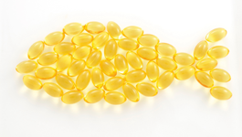 We're always being told fish oil is good for us. Eat more oily fish. Well apparently there is accumulating evidence, according to S. J. Gladman, that fish oils, and in particular a class of fish oils known as Omega-3, has some remarkable therapeutic potential in a number of disorders of the nervous system, including trauma. Using a cell-based model to replicate the mechanical stretch axons experience around the injury site of a cord lesion, the group from London, who are developing this concept and hope to take it to clinical trial in the near future, have shown significant reduction in cell death after treatment with Omega-3. They went further and found that if, in this model, they used neuronal cells derived from mice that have naturally high levels of Omega-3 AND added Omega-3 cell death was totally abolished. Omega-3 appears to confer neuroprotective effects and will likely only be useful if given in the immediate aftermath of injury.
[text added to post]
Omega-3 polyunsaturated fatty acids have different chemical forms, for example docosahexaenoic acid (DHA) or alpha-linolenic acid (ALA). Jodie Hall presented a poster on the effects on dietary eicosapentaenoic acid (EPA) in rats with SCI. Surprisingly, when given an EPA-rich diet rats did less well in locomotors tests in contrast to when they received EPA by injection. Why this is is unclear and further work is ongoing to understand why the route of delivery is so important.
Elsewhere M. S. Joseph combined Omega-3 with curcumin (a curry spice) in the diet and found it enhanced learning of new tasks by circuits in the spinal cord after SCI. I suppose we should really be eating fish curry.
Subscribe to:
Posts (Atom)

 Posts
Posts

